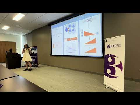
This research investigates how glutamine-rich regions within the LAG-3 protein influence Notch signaling, a critical pathway for cell communication and development. Using CRISPR gene editing, the study found that removing glutamine repeats alters stem cell behavior and cell-cycle progression, providing insights relevant to cancer, Alzheimer’s disease, and future therapies.

This research investigates how the olfactory system of the Spanish ribbed newt adapts between aquatic and terrestrial environments. By analyzing cellular and genetic changes in the nose, the study reveals remarkable sensory plasticity, offering broader insights into nervous system flexibility and potential implications for understanding neurodegenerative diseases such as dementia.
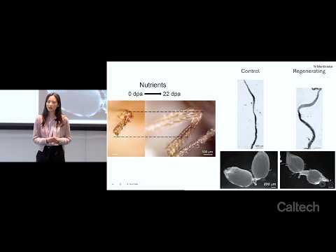
This research investigates whether regeneration can be induced in animals that normally lack regenerative abilities. Using nutrient factors such as amino acids and insulin, regeneration was stimulated in mice, jellyfish, and fruit flies. The findings reveal that regeneration is a coordinated whole-body process involving energy allocation, organ remodeling, and conserved nutrient signaling pathways.

This research investigates how coordinated and random patterns of cell division shape facial development. Using 3D imaging of mouse embryos, it reveals that the direction of cell division—not just its location—drives tissue growth. Balancing orderly and chaotic cellular behaviours may be key to understanding healthy face formation and preventing developmental defects.
2025
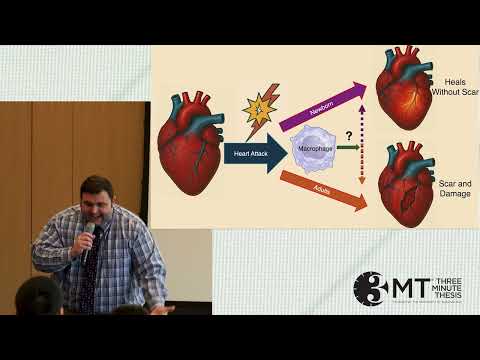
This research explores how tissue-resident macrophages guide immature heart muscle cells during early development. By identifying immune signals that enable scar-free heart regeneration in newborns, the work aims to uncover therapeutic pathways that could restore regenerative capacity and improve outcomes for patients with heart disease.
Craniosynostosis occurs when skull sutures fuse too early, requiring risky surgeries. The researcher identified microRNA-200A as a key regulator of suture development. In mice lacking miR-200A, sutures fused prematurely, but adding extra miR-200A via gene therapy prevented fusion entirely. This breakthrough suggests a non-surgical future treatment for craniosynostosis.

Cleft lip formation may result from broken DNA enhancers—switches that control facial development genes. Scanning the genomes of 130 African children with clefts, this research identified harmful enhancer variants and confirmed their effects in mouse models. The disrupted enhancer likely regulates BMP2, offering new insight into cleft biology and future prevention.

Balanced cell growth is essential: too much can cause cancer, too little can cause skeletal disorders. This PhD project investigates a mysterious protein linked to dwarfism. By tagging it with GFP, the researcher discovered it drives fat-droplet formation, revealing a previously unknown function that may explain its powerful effects on body growth.